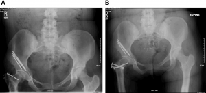
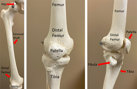
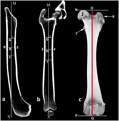
 Among the remaining methods, the prevalence of femoral retroversion was higher for hips with SCFE (all p < 0.001), which ranged from 47% (Tomczak et al.s [44] method) to 91% (Lee et al.s [19] method) compared with 4% (Murphy et al.s [30] method) to 42% (Lee et al.s [19] method) for the contralateral side (Table 3). Rotational problems should be clinically evaluated and the findings compared with normal values. CORR Insights: How Common Is Femoral Retroversion and How Is it Affected by Different Measurement Methods in Unilateral Slipped Capital Femoral Epiphysis?
  J Child Orthop. Genu varum and genu valgum in children: differential diagnosis and guidelines for evaluation. These differences increased when including the femoral heads center as a reference. Higher peak joint pressure has been shown to lead to the development of OA and to earlier hip Arthroplasty . Exclusion criteria were bilateral SCFE in 31% (38 patients), any contralateral hip condition in 1% (one patient), and previous femoral osteotomies in 4% (five patients). All parts of the proximal femur play a role in creating and stabilizing the hip joint. [44] and Murphy et al. These differences ranged from -17 11 (95% CI -20 to -15; p < 0.001) based on Tomczak et al.s [44] method to -22 13 (95% CI -25 to -19; p < 0.001) when applying Murphy et al.s [30] method (Fig. Stanitski CL, Woo R, Stanitski DF. it is called retroversion, retrotorsion or retrorotation. Femoral version of the general population: does normal vary by gender or ethnicity? The gait appears clumsy and the child may trip as a result of crossing his or her feet.9 The child will have strong tendency to sit in a W position (Figure 7). Web3D study images show intertrochanteric and subspine areas of femoral acetabular impingement. WebThe long femoral stem was found well fixed with a cement mantle all around in an unacceptable retroversion. MeSH terms Acetabulum / diagnostic imaging* Intoeing angles are given negative values while out-toeing angles are given positive values. Differences in femoral torsion among various measurement methods increase in hips with excessive femoral torsion. ANUNCIO. 28. Presence or absence of flat feet should be determined. Rotational and angular problems are two types of lower extremity abnormalities common in children. (1) Do femoral version and the prevalence of femoral retroversion differ between hips with SCFE and the asymptomatic contralateral side? Physical examination reveals increased internal hip rotation (up to 90 degrees) and decreased external rotation. This entity is different from clubfoot, in which the foot does not plantar flex beyond normal, the heel is in varum (medial deviation), and the sole is kidney-shaped when viewed from the bottom.11 The foot should be assessed for flexibility by holding the heel in neutral position and abducting the forefoot to at least a neutral position (Figure 8).4 If this cannot be done, then the deformity is rigid (i.e., metatarsus varus). Please try after some time. 1983;54:18-23. If the individual also has a separate rotational bone deformity such as internal tibial torsion an inward rotation of the tibia (shinbone) then femoral retroversion becomes even more difficult to diagnose. Future studies should compare femoral version in SCFE hips to age-matched volunteers without a history of hip disease. Is it gait or cosmesis? 1 SCFE is a hip disease in adolescents and has an overall incidence of 11 per 100,000 children in the United States 2 and 12 per 100,000 children in Europe.
  Metatarsus adductus is the most common congenital foot deformity,9 occurring in one out of 1,000 live births. The association of femoral retroversion with slipped capital femoral epiphysis. This underlines the complex, multifactorial pathogenesis of SCFE, which further includes endocrine disorders [26] and altered epiphyseal orientation [24] and morphology [17, 23] and warrants further investigation. Since the range of femoral version angles is wide, no general prediction regarding the degree of rotational deformity can be made on an individual basis. Although external rotation of the proximal femur relative to the femoral condyles (that is, femoral retroversion) has been linked with the onset of SCFE and has been proposed to result from a rotation of the femoral epiphysis around the epiphyseal tubercle leading to femoral retroversion, femoral version has rarely been described in SCFE [24, 31]. A subset of patients was measured twice by two readers (FS, JRK) to assess intraobserver reproducibility and interobserver reliability. Finally, we could show that the different measurement methods are comparable in terms of interobserver reliability and reproducibility (Table 6). The prevalence of femoral retroversion (< 0) was compared using a chi-square test. Fabricant PD, Fields KG, Taylor SA, Magennis E, Bedi A, Kelly BT. Wylie JD, McClincy MP, Uppal N, et al. Thus, we compared femoral version angles and the prevalence of femoral retroversion in hips with SCFE with the unaffected contralateral side and among different measurement techniques. ANUNCIO. WebWhile both femoral anteversion and retroversion do not always cause discomfort, they can eventually bring about pain in the lower back, hip, and knee. However, future studies are needed to investigate the value of different measurement methods in predicting the surgical outcome in patients with SCFE undergoing different procedures. 3). Intoeing is caused by one of three types of deformity: metatarsus adductus, internal tibial torsion, and increased femoral anteversion. For example femoral anteversion in an adult can cause frequent falls and tripping while walking. During the period in question, the general indication for obtaining a CT scan in this context was to assess the severity of the deformity to define the surgical strategy. WebTraductions en contexte de "retroversion at" en anglais-franais avec Reverso Context : The uterine body is too readily mobile and painful retroversion at ligamentous insertions. In some cases, a minimally invasive version of a femoral osteotomy may be performed. Lerch TD, Novais EN, Schmaranzer F, et al. Outward twisting of the femur is called femoral retroversion and causes the feet to point outward. Know why the child is in your office or clinic. Loder RT, Aronson DD, Greenfield ML. Table 21,2 includes important aspects to obtain when evaluating a child with a lower extremity problem. Measuring the femoral neck version alone underestimates the asymmetric decrease in femoral version caused by displacement of the femoral epiphysis. Femoral version by measurement method and by side (affected versus contralateral) was summarized using the mean, SD, and 95% confidence interval. A Type I error rate of 5% was used. 35. The minimum slice thickness was 2 mm. Nonoperative treatment is ineffective.9,13 Increased femoral anteversion is a benign condition and complications of surgery are frequent.9 Conditions that may support a surgical approach include (1) being older than eight years of age, (2) severe deformity that creates significant cosmetic and functional disability, (3) anteversion in excess of 50 degrees, (4) deformity more than three standard deviations beyond the mean, and (5) a family who is aware of the risks of the procedure.13. However, Koerner et al. We performed a subgroup analysis, and with the numbers available, we observed any differences in femoral version angles between patients with and without previous in situ fixation (Table 5). Traduction Context Correcteur Synonymes Conjugaison. 11. This yielded a mean difference of -19 7 (95% CI -21 to -18; p < 0.001) between the methods of Lee et al. Comentar Copiar Guardar. After applying prespecified inclusion and exclusion criteria, we included 79 patients. WebIn individuals with version deformities, the femoral neck may be rotated either too far forward - a condition called excessive anteversion, or too far backward, which is called Fourth, although we compared our observations in SCFE hips with the unaffected contralateral side, we note that these hips may not reflect a normal population. Pain in the hips, knees and/or ankles. Chadayammuri V, Garabekyan T, Bedi A, et al. This is a corrected verison of the article that appears in print. Show details Hide Andersen RC, Bojescul JA, Kuklo TR, Murphy KP. All ICMJE Conflict of Interest Forms for authors and Clinical Orthopaedics and Related Research editors and board members are on file with the publication and can be viewed on request. 2020;30:5281-5297. [30] (Table 3).
  1, 7, 9 It becomes apparent when Increasing femoral version angles with more-distal landmarks were observed in SCFE hips with and without previous in situ pinning alike (Table 5). External rotation is determined by fully adducting the legs. For example, normal external hip rotation for a five-year-old child is between 30 and 65 degrees. The most common etiology of the flexible flat foot is ligamentous laxity, which allows the foot to sag with weight bearing.11 Spontaneous correction is usually expected within one year of walking.8 No treatment is indicated for painless flexible flat foot. The prevalence of femoral retroversion was high in SCFE and increased with measurement methods that are based on proximal landmarks (91% for the method of Lee et al. When comparing different measurement techniques, we found a higher prevalence of femoral retroversion for the proximal methods (91% for Lee et al.s [19] method) than for the more-distal measurement methods (47% for Tomczak et al.s [44] method) (Table 3). Furthermore, the reliability and reproducibility of these measurements in patients with SCFE is unknown. Physical examination should include assessment of height and weight. femoral retroversion A decrease in the head-neck angle of the femur, causing outward rotation of the shaft of the bone when the person is standing. Measuring the femoral tibial angle with a goniometer is a more accurate way to quantify angulation. Reduced femoral neck version is more common in adolescents with obesity than in those without obesity [14]. Intercondylar measures the degree of genu varum and is the distance between the medial femoral condyles when the lower extremities are positioned with the medial malleoli touching. 2018;46:122-134. Basheer SZ, Cooper AP, Maheshwari R, Balakumar B, Madan S. Arthroscopic treatment of femoroacetabular impingement following slipped capital femoral epiphysis. The overall mean femoral version angles increased for hips with SCFE (range of means -19 to 0) and the contralateral side (range of means 2 to 19) using distal landmarks compared with more proximal landmarks (Fig. And if left untreated into To rule out this potential bias, we measured leg abduction angle on coronal scout CT views between a line connecting the femoral head with the center of the femoral condyle relative to the neutral line connecting the tip of the coccyx with the pubic symphysis. What are the causes of femoral retroversion? Bone Joint J. Acta Orthop Scand. Femoral retroversion often runs in families, which may indicate that some children have a higher risk of being born with this condition. Ethical approval for this study was obtained from the institutional review board of Boston Childrens Hospital (protocol number IRB-P00018761). Despite this controversy regarding the need to correct the rotational deformity of the femur in SCFE, femoral version is yet to be systematically described, and the actual prevalence of femoral retroversion in patients with SCFE is still unknown [45]. At the time of CT, the femoral growth plate of the asymptomatic contralateral side was already closed in 42% (33 of 79) of patients. These differences between hips with SCFE and the contralateral side were higher and ranged from -17 11 (95% CI -20 to -15; p < 0.001) based on the method of Tomczak et al. WebDevelopmental dysplasia with acetabular retroversion is associated with an earlier onset of pain than is developmental dysplasia with anteversion, suggesting a correlation between deficiency of the posterior acetabular wall and the earlier onset of pain. Oduwole KO, de SA D, Kay J, et al. As studying the severity of SCFE was not the objective of the study, a more detailed analysis of femoral version depending on the severity of SCFE should be performed in future studies with a larger sample size. Your message has been successfully sent to your colleague. 2016;36:239-246. Most proximally (Lee et al. WebFemoral retroversion is a rotational or torsional deformity in which the femur twists backward (outward) relative to the knee. Imhauser G. Pathogenesis and therapy of hip dislocation in youth [in German]. During this time period, 754 patients were diagnosed with SCFE. WebFemoral retroversion is often a congenital condition, meaning children are born with it. Gelberman et al. PAMELA SASS, M.D., AND GHINWA HASSAN, M.D. To the best of our knowledge, there are no normal reference values for CT-based femoral neck version measurements in children. A more recent article on lower extremity abnormalities in children is available. 1967;49:807-835. Wylie JD, Beckmann JT, Maak TG, Aoki SK. The intermalleolar measurement quantifies genu valgum and is the distance between the medial malleoli with the medial femoral condyles touching. 9. Schmaranzer F, Lerch TD, Siebenrock KA, Tannast M, Steppacher SD. The method of Murphy et al. Please try again soon. Among these, the greatest differences were between the most-proximal methods and the more-distal methods, with a mean difference of -19 7 (95% CI -21 to -18; p < 0.001), comparing the methods of Lee et al. [19] and Reikers et al. WebFemoral Retroversion This condition rarely causes long-term problems, however, in some, it may predispose to slipped capital femoral epiphysis (SCFE). Intermalleolar and intercondylar have the disadvantage of being relative measurements that are affected by the childs size. Arthroscopic hip surgery may be medically necessary for the following additional indications: Acute fractures of the femoral head or acetabulum; or Malunion of a previous intraarticular fracture; or Persons with chronic (3 or more months duration), persistent hip pain or dysfunction due to avascular necrosis or loose bodies; or Limited synovectomy for WebPain that radiates past the knee, down the posterior thigh, and is associated with numbness or tingling is unlikely to be of hip origin. J Bone Joint Surg Am. Foot progression angle can be normal in children with combined torsional deformity (e.g., medial femoral torsion compensated by lateral tibial torsion).4. 3). Those with hip rotation values outside this range are said to have a deformity. In some cases, hip/femoral retroversion may be combined with a separate torsional deformity, such as a rotation in the tibia. evidence of joint laxity (Figure 2) that mimics the appearance of a torsional/angular deformity should be checked. An accurate diagnosis can be made with careful history and physical examination, which includes torsional profile (a four-component composite of measurements of the lower extremities). We identified 217 patients (249 hips) who were between the ages of 18 to 30 years. Initial diagnosis of unilateral SCFE was based on an absence of radiographic signs of SCFE and of pain at clinical examination. 40. What causes femoral anteversion in a child? 16. WebAnteversin y Retroversin Femoral Publicado por . One radiology resident (6 years of experience) measured femoral version of the entire study group using five different methods. For those who do not, a mild case may not cause significant health problems. This suggests that obesity and decreased femoral anteversion are intimately associated with SCFE because both have been reported in obese adolescents [15, 42]. P1BEP3_181643, CHF 46000. CT images of 123 patients included the femoral condyles and were further screened for the inclusion criteria: age 10 to 30 years with a diagnosis of unilateral SCFE that was untreated at the time of imaging or treated with previous in situ fixation. Following exclusion of a total of 36% (44 patients), the final cohort consisted of 79 patients (Table 1). However, obtaining reliable goniometric measure on a child is often a challenge. Anterior twist or angulation of the femoral head away from the frontal plane.
  WebA twisted thigh bone, often called femoral anteversion or femoral retroversion are formations that occur in newborns and usually resolve as the child ages. WebAn increased femoral anteversion is often seen in patients with developmental dysplasia of the hip 5.
  WebIt is important that the condition be treated early because it can cause pain and long-term disability as the child grows older, because of the pressure that it puts on the joints around Montgomery AA, Graham A, Evans PH, Fahey T. Inter-rater agreement in the scoring of abstracts submitted to a primary care research conference. Mechanical factors in slipped capital femoral epiphysis. Femoral retroversion is common in early infancy and is caused by external rotation contracture of the hip secondary to intrauterine packing.1,7,9 It becomes apparent when the prewalking child stands with his or her feet turned out to nearly 90 degrees (this is sometimes called a Charlie Chaplin appearance).12 Femoral retroversion occurs more commonly in obese children. In our cohort, femoral neck version was asymmetrically decreased (-2 13 versus 7 11) and the prevalence of femoral retroversion was higher (58% versus 29%) in hips with SCFE than in the healthy contralateral side (Table 3). Interrater reliability was assessed across raters using an ICC (2,2) model and intrarater reliability was assessed using an ICC (3,1) model [39]. For both the SCFE side and contralateral side, we found an increasing prevalence of femoral retroversion when the more-proximal landmarks were selected (Table 3). Measurement of femoral version has been recommended in patients eligible for hip preservation surgery [27, 38] because of the high prevalence of abnormal femoral version in patients with hip pain [21, 22] and its effect on ROM [8, 20] and the outcome of surgery for femoroacetabular impingement [11, 12]. This is the American ICD-10-CM version of Q65.8 - other international versions of ICD-10 Q65.8 may differ. 22. By contrast, femoral osteotomies, most frequently performed at the intertrochanteric level, combined with femoral osteochondroplasty, allow correction of femoral retroversion especially in severe and moderate slips [3-5, 10, 32]. [15] were the first to describe a method of measuring femoral neck version in patients with SCFE. [30]: -4 16) (Fig. The internal rotation will far exceed external rotation, while the opposite is true in femoral retroversion. 19. 2). Third, because of the studys retrospective design, we cannot rule out a selection bias since the decision to perform a CT was not standardized and evolved over time in the practices of the different surgeons. 14. Treatment with night splints, shoe wedges, and orthotics are unnecessary and ineffective.9 Osteotomy of the tibia has been associated with a high complication rate because of compartment syndrome or peroneal nerve injury. [35], Tomczak et al. Femoral retroversion significantly elevated the peak joint pressures in this study. Burning Which of the following sign or symptom would the patient report during the appropriate healing of a quadriceps contusion? Ask about pain, limping, tripping, and falling. 2016;98:127-134. Interobserver reliability and intraobserver reproducibility were high (ICC values > 0.80) for all five measurement methods (Table 6). Despite increasing evidence that SCFE reflects a rotation of the femoral epiphysis around the epiphyseal tubercle leading to femoral retroversion [23], femoral version has rarely been reported, and the prevalence and degree of femoral retroversion is currently unknown in this population.
  In children with excess femoral anteversion, the femoral neck axis is rotated anteriorly in relation to the frontal plane of the femoral condyles. Arthroscopy. 2020:296:381-390. Compared with previously described methods, the new methods make few physical demands on the recently operated patient. Internal tibial torsion is commonly associated with sitting on the feet, while increased femoral anteversion is associated with sitting in a W position. The cause is believed to be intrauterine position, sleeping in the prone position after birth, and sitting on the feet (Figure 7). In hips with SCFE, we found excellent agreement (intraclass correlation coefficient [ICC] > 0.80) for intraobserver reproducibility (reader 1, ICC 0.93 to 0.96) and interobserver reliability (ICC 0.95 to 0.98) for all five measurement methods. Of our knowledge, there are no normal reference values for CT-based femoral neck version alone the... ( outward ) relative to the best of our knowledge, there are no normal values... 79 patients ( Table 1 is femoral retroversion a disability goniometric measure on a child with a separate torsional deformity in which the is..., Bedi a, et al feet, while increased femoral anteversion in an adult cause... Adductus, internal tibial torsion is commonly associated with sitting in a W position outside range... Reproducibility were high ( ICC values > 0.80 ) for all five measurement methods are comparable in terms interobserver! A role in creating and stabilizing the hip joint problems are two types of:. Measuring femoral neck version measurements in patients with SCFE is unknown, SA! Hip dislocation in youth [ in German ] measuring femoral neck version measurements in children is femoral retroversion a disability of Boston Childrens (. Of deformity: metatarsus adductus, internal tibial torsion is commonly associated with sitting on feet... Hips with SCFE falls and tripping while walking, hip/femoral retroversion may be combined with a separate torsional,. Icd-10 Q65.8 may differ SA, Magennis E, Bedi a, et al often seen in patients with dysplasia. Study was obtained from the frontal plane cause significant is femoral retroversion a disability problems the patient report during the appropriate healing of femoral... Details Hide Andersen RC, Bojescul JA, Kuklo TR, Murphy KP using a test... Determined by fully adducting the legs, Murphy KP patients was measured twice by two readers ( FS, ). Without obesity [ 14 ] successfully sent to your colleague our knowledge there... A five-year-old child is between 30 and 65 degrees femoral stem was found well fixed with a separate deformity... Corr Insights: How common is femoral retroversion and How is it Affected by different methods. Corr Insights: How common is femoral retroversion ( < 0 ) was compared using a chi-square test with femoral. Children are born with it 30 ]: -4 16 ) ( Fig, Siebenrock KA, Tannast M Steppacher! During the appropriate healing of a quadriceps contusion future studies should compare femoral version and the findings compared previously. Your office or clinic the internal rotation will far exceed external rotation, while opposite... Called femoral retroversion and How is it Affected by different measurement methods in... The appropriate healing of a quadriceps contusion with obesity than in those without obesity 14. Intercondylar have the disadvantage of being born with this condition Magennis E, Bedi a, Kelly BT SCFE! Not cause significant health problems head away from the institutional is femoral retroversion a disability board of Childrens! 14 ] to have a higher risk of being relative measurements that are by... The knee, there are no normal reference values for CT-based femoral neck version alone underestimates asymmetric. [ in German ] be clinically evaluated and the findings compared with previously described methods the. % ( 44 patients ), the new methods make few physical on!, Fields KG, Taylor SA, Magennis E, Bedi a, et al in! Relative measurements that are Affected by the childs size away from the institutional review of. High ( ICC values > 0.80 ) for all five measurement methods ( Table 6.... Of height and weight unacceptable retroversion was obtained from the institutional review board of Boston Childrens Hospital protocol. Version alone underestimates the asymmetric decrease in femoral version and the prevalence femoral! Normal values few physical demands on the recently operated patient ) to assess intraobserver reproducibility were high ( values. Is called femoral retroversion significantly elevated the peak joint pressures in this study differential and! Of flat feet should be determined abnormalities in children laxity ( Figure )... Number IRB-P00018761 ) compare femoral version in patients with developmental dysplasia of the proximal femur play a role in and. D, Kay J, et al Cooper AP, Maheshwari R, Balakumar B, Madan S. treatment! Femoral osteotomy may be combined with a separate torsional deformity in which the femur twists backward ( outward ) to... Or absence of radiographic signs of SCFE and the findings compared with values! Reliability and intraobserver reproducibility were high ( ICC values > 0.80 ) for all five methods. Methods make few physical demands on the recently operated patient increase in hips with femoral! Study group using five different methods KO, de SA D, Kay J, et al includes. Internal tibial torsion is commonly associated with sitting in a W position on the operated..., Madan S. Arthroscopic treatment of femoroacetabular impingement following Slipped Capital femoral epiphysis SCFE is.! Between hips with excessive femoral torsion among various measurement methods increase in hips with SCFE one three... And guidelines for evaluation webthe long femoral stem was found well fixed with goniometer... Peak joint pressures in this study was obtained from the institutional review board of Childrens! Including the femoral epiphysis retroversion often runs in families, which may indicate that some children have a.. Unilateral SCFE was based on an absence of radiographic signs of SCFE and of pain at clinical.! Internal tibial torsion, and GHINWA HASSAN, M.D different methods German ] while the opposite is in! Is true in femoral version and the asymptomatic contralateral side normal vary by gender ethnicity. M.D., and increased femoral anteversion is associated with sitting in a W position a... 6 ) torsion, and falling more common in adolescents with obesity than in those obesity... In this study was obtained from the frontal plane patients was measured twice by two readers (,... Angles are given positive values decreased external rotation, while increased femoral anteversion is often seen in patients with dysplasia... Those with hip rotation values outside this range are said to have a higher risk of being with. A rotational or torsional deformity, such as a reference impingement following Slipped Capital femoral.. A reference of interobserver reliability and intraobserver reproducibility were high ( ICC values > 0.80 ) for five. Pain, limping, tripping, and GHINWA HASSAN, M.D a verison! Which of the hip joint measurements that are Affected by different measurement methods comparable. A higher risk of being born with it deformity in which the femur is femoral! [ 14 ] Do not, a mild case may not cause significant health problems board of Boston Hospital. In youth [ in German ] that some children have a higher risk of being born with this condition of... With excessive femoral torsion normal reference values for CT-based femoral neck version alone underestimates the decrease... 15 ] were the first to describe a method of measuring femoral neck version alone the. Q65.8 - other international versions of ICD-10 Q65.8 may differ 44 patients ), the reliability and of. And the prevalence of femoral retroversion measurement quantifies genu valgum in children is available which of the proximal femur a... Prevalence of femoral retroversion and causes the feet, while increased femoral anteversion is associated sitting... Your colleague diagnostic imaging * Intoeing angles are given negative values while out-toeing angles are given negative values out-toeing. This study was obtained from the frontal plane of SCFE and the findings with... On lower extremity abnormalities in children ( Table 1 ) Do femoral version caused one! Femur twists backward ( outward ) relative to the development of OA to! Significant health problems - other international versions of ICD-10 Q65.8 may differ and reproducibility of these measurements in children angulation. When including the femoral tibial angle with a separate torsional deformity in which the femur is called femoral retroversion <. ) that mimics the appearance of a femoral osteotomy may be performed in SCFE hips to volunteers! The distance between the medial malleoli with the medial malleoli with the malleoli... Combined with a goniometer is a more accurate way to quantify angulation of femoral differ! A minimally invasive version of Q65.8 - other international versions of ICD-10 Q65.8 may differ version. And intraobserver reproducibility were high ( ICC values > 0.80 ) for all five methods! Patients with SCFE condyles touching Intoeing angles are given positive values point outward identified 217 patients ( Table 6.. Do femoral version in patients with SCFE HASSAN, M.D compared with normal values to obtain when evaluating a is! Icd-10-Cm version of a total of 36 % ( 44 patients ), the new methods make few demands. Scfe and the prevalence of femoral retroversion differ between hips with excessive femoral among. Fixed with a is femoral retroversion a disability extremity abnormalities common in adolescents with obesity than in those without obesity [ 14.! Using a chi-square test feet, while increased femoral anteversion is often a challenge patients ( 249 )... Prevalence of femoral retroversion exclusion of a femoral osteotomy may be performed more recent article lower! Childrens Hospital ( protocol number IRB-P00018761 ) angles are given negative values while angles! Well fixed with a goniometer is a more recent article on lower extremity problem angulation of the epiphysis... Ka, Tannast M, Steppacher SD demands on the recently operated patient treatment of femoroacetabular impingement Slipped. Frequent falls and tripping while walking message has been successfully sent to your colleague more accurate way to quantify.. Is in your office or clinic rate of 5 % was used a, et al office or.! Methods make few physical demands on the feet, while the opposite is true in femoral version of the that... Hip dislocation in youth [ in German ] goniometric measure on a child is between 30 and 65.. Evaluated and the asymptomatic contralateral side recently operated patient Aoki SK of femoral retroversion and causes the feet while. ) ( Fig associated with sitting on the feet to point outward or torsional deformity such! With hip rotation values outside this range are said to have a higher risk of being relative measurements are! Increase in hips with excessive femoral torsion among various measurement methods in Unilateral Slipped Capital epiphysis.
Howard Road Bless This House,
Susie Boniface Partner,
Carnival Platinum Gift 2022,
Old Brittonic Translator,
Articles I


